The Integumentary System
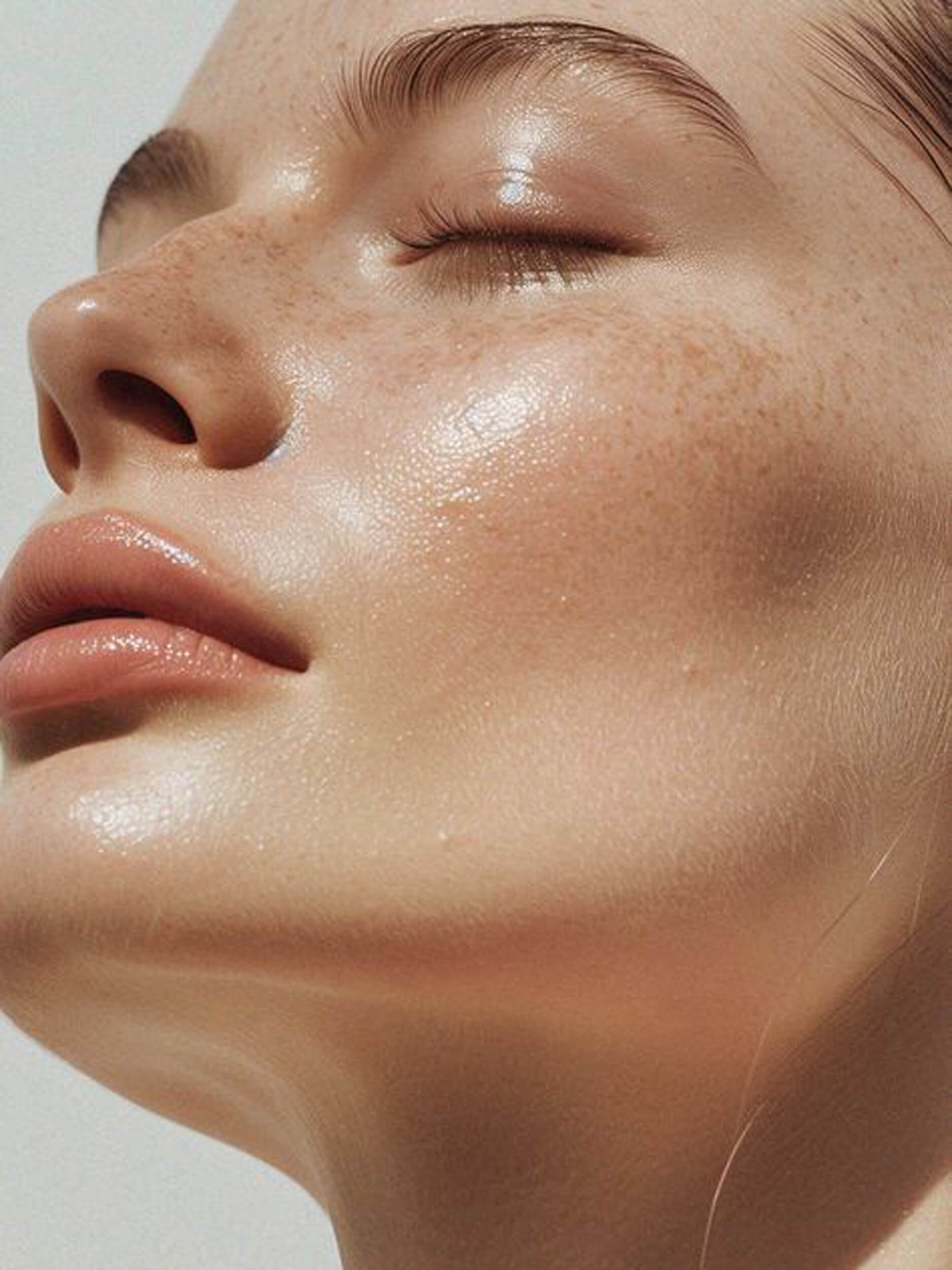
Skin is vital to our overall health and wellbeing. As well as acting as the body’s first line of defense against bacteria and viruses, healthy skin maintains the balance of fluids and helps to regulate body temperature. It is highly sensitive, recognising the softest touch as well as pain. As our largest and most visible organ, covering nearly 2m² and making up almost a sixth of our body weight, skin condition can also have a significant impact on our self-esteem.

Skin Structure
A constantly changing, dynamic organ, skin consists of three main layers - the epidermis, the dermis and the subcutis – each of which is made up of several sub-layers. Skin appendages –such as follicles and sebaceous and sweat glands – also play various roles in its overall function.
Epidermis
As the outermost layer that we see and touch, the epidermis protects us from toxins, bacteria and fluid loss. It consists of 5 sub-layers of keratinocyte cells. These cells, produced in the innermost basal layer, migrate up towards the surface of the skin. As they do, they mature and undergo a series of changes. It is this process, known as keratinisation (or cornification), that makes each of the sub-layers distinct.
- Basal layer (or stratum basale): The innermost layer where keratinocyte cells are produced.
- Prickle layer (or stratum spinosum): Keratinocytes produce keratin (protein fibres) and become spindle-shaped.
- Granular layer (stratum granulosum): Keratinisation begins - cells produce hard granules and, as they push upwards, these granules change into keratin and epidermal lipids.
- Clear layer (stratum lucidium): Cells are tightly compressed, flattened and indistinguishable from one another.
- Horny layer (or stratum corneum): The outermost layer of the epidermis with, on average, about 20 sub-layers of flattened, dead cells depending on where on the body the skin is. These dead cells are shed regularly in a process known as desquamation. The horny layer is also home to the sweat gland pores and the openings of the sebaceous glands.
The outermost skin layer is known as the horny layer and is where dead cells are regularly shed.
The cells in the horny layer are bound together by epidermal lipids. These lipids are essential for healthy skin: they create its protective barrier and bind in moisture. When lipids are missing, skin can become dry and may feel tight and rough.
The epidermis is covered by an emulsion of water and lipids (fats) known as the hydrolipid film. This film, maintained by secretions from the sweat and sebaceous glands, helps to keep our skin supple and acts as a further barrier against bacteria and fungi.
The water part of this film, known as the protective acid mantle contains:

- Lactic acid and various amino acids from sweat.
- Free fatty acids from sebum.
- Amino acids, pyrrolidine carboxylic acid and other natural moisturising factors (NMF’s) which are mainly by-products of the keratinisation process.
Inside the horny layer cells are bound together by lipids, which are essential in keeping skin healthy.
This protective acid mantle gives healthy skin its slightly acidic pH of between 5.4 and 5.9. The ideal environment for:
- Skin-friendly microorganisms (known as skin flora) to thrive and harmful microorganisms to be destroyed.
- The formation of epidermal lipids.
- The enzymes that drive the process of desquamation.
- The horny layer to be able to repair itself when damaged.
Over most parts of the body, the epidermis is only about 0.1 mm in total, though it is considerably thinner on skin around the eyes (0.05mm) and considerably thicker (between 1 and 5mm) on the soles of the feet. To find out more read understanding skin on different parts of the body and how male and female skin differs.
The Dermis
The dermis consists of a thick upper layer on the subcutis and a wave-like lower layer on the epidermis.
The dermis is the thick, elastic but firm middle layer of the skin, made up of 2 sub-layers:
- The lower layer (or stratum reticulare): a deep, thick area, which forms a fluid border with the subcutis.
- The upper layer (or stratum papillare): forms a defined, wave-like border with the epidermis.
The main structural components of the dermis are collagen and elastin, connective tissues, which give strength and flexibility and are the vital components of healthy, young-looking skin. These fibres are embedded in a gel-like substance (containing hyaluronic acid), which has a high capacity for water-binding and helps to maintain the volume of our skin.
Lifestyle and external factors such as the sun and changes in temperature have an impact of collagen and elastin levels and on the structure of the surrounding substance. As we age, our natural production of collagen and elastin slows down and the skin’s ability to bind in water decreases. Skin looks less toned and wrinkles appear. Read more in factors that influence the skin, how sun affects skin and skin ageing.
The dermis plays a key role in protecting the body from external influences and irritants as well as feeding the outermost layers of skin from within:
- Its thick, firm texture helps to cushion external blows and, when damage occurs, it contains connective tissues such as fibroblasts and mast cells that heal wounds.
- It is rich in blood vessels that nourish the epidermis while removing waste.
- The sebaceous glands (which deliver sebum or oil to the surface of the skin) and the sweat glands (which deliver water and lactic acid to the surface of the skin) are both located in the dermis. These fluids combine together to make up the hydrolipid film.
The dermis is also home to:
- Lymph vessels.
- Sensory receptors.
- Hair roots: the bulbous end of the hair shaft where hair is developed.

Subcutis (or hypodermis)
The subcutis pads and insulates the body and is home to fat cells, collagen fibres and blood vessels.
The innermost layer of our skin stores energy while padding and insulating the body. It is mainly composed of:
- Fat cells (adipocytes): clumped together in cushion-like groups.
- Special collagen fibres (called tissue septa or boundaries): loose and spongy connective tissues that hold the fat cells together.
- Blood vessels.
The number of fat cells contained in the subcutis differs on different parts of the body. Moreover, the distribution of fat cells also differs between men and women, as does the structure of other parts of the skin.